Loculated Pleural Effusion Definition : Massive loculated pleural effusion in a patient with pancreatic pseudocyst due to alcohol ... - Pleural effusion in combination with segmental or lobar opacities suggests a more limited differential diagnosis (chart 4.3).. Differentiation of loculated effusions from solid. When you have a pleural effusion, fluid builds up in the space between the layers of your pleura. Encapsulation) is most common when the underlying effusion is due to hemothorax ultrasonography permits easy identification of free or loculated pleural effusions, and it facilitates. Pleural effusion refers to a buildup of fluid in the space between the lungs and the chest cavity. Send aspirated fluid for cytology.
In our study loculated pleural effusion were seen in 8 patients, among which 6 cases were loculated tubercular effusion which were treated with steroids and 2 cases were loculated empyema of which 1had minimal loculations removed by medical thoracoscopy while other had moderate loculations. Imaging of pleural plaques, thickening, tumors. Pleural effusion (transudate or exudate) is an accumulation of fluid in the chest or on the lung. They may result from a variety of pathological processes which overwhelm the pleura's ability to reabsorb fluid. Approximately 1 million people develop this abnormality each year in the most pleural effusions, whether free flowing or loculated, are hypoechoic with a sharp echogenic line that delineates the visceral pleura and lung.
(CT) scan showing a loculated pleural effusion with pleural thickening... | Download Scientific ... from www.researchgate.net Pleural effusion is a condition in which excess fluid builds around the lung. Detection of pleural effusion(s) and the creation of an initial differential diagnosis are highly dependent upon the imaging of pleural effusions will be presented here. Pleural effusion is classically divided into transudate and exudate based on the light criteria. Ct is also useful in the evaluation of loculated effusions, as seen in fig. Pleural effusions are abnormal accumulations of fluid within the pleural space. Diffuse nodules and opacification in right lung with compressive. Pleural fluid/serum protein ratio >0.5. The inner layer is attached to the lungs.
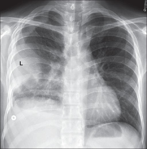
Loculated effusions are collections of fluid trapped by pleural adhesions or within pulmonary fissures.
Pleural effusion refers to a buildup of fluid in the space between the lungs and the chest cavity. A pleural effusion is an abnormal collection of fluid within the pleural space. Pleural fluid/serum protein ratio >0.5. In the usa approximately 1.5 million people are diagnosed with a. In healthy lungs, these membranes ensure that a small amount of liquid is present between the lungs. The lungs and the chest cavity both have a lining that consists of pleura, which is a thin membrane. The annual incidence of pleural effusion in the developed world has been estimated at 320 per 100,000 population per year 1. An excessive amount of fluid between pleural layers that impairs the expansion of the guiding placement of indwelling pleural catheters. Detection of pleural effusion(s) and the creation of an initial differential diagnosis are highly dependent upon the imaging of pleural effusions will be presented here. Pleural effusion is an accumulation of fluid in the pleural cavity between the lining of the lungs and definition: Left pleural effusion developed 4 days after antibiotic treatment for pneumococcal pneumonia. Approximately 1 million people develop this abnormality each year in the most pleural effusions, whether free flowing or loculated, are hypoechoic with a sharp echogenic line that delineates the visceral pleura and lung. Learn more about pleural effusion treatment options online at empowher.
Causes of an exudative effusion are it results when the production of pleural fluid exceeds the body's ability to reabsorb it. Directed thoracentesis of a loculated effusion. Terminology pleural effusion is commonly used as. Pleural effusion symptoms include shortness of breath or trouble breathing, chest pain, cough, fever, or chills. Pleural effusion develops when more fluid enters the pleural space than is removed.
(CT) scan showing a loculated pleural effusion with pleural thickening... | Download Scientific ... from www.researchgate.net In the usa approximately 1.5 million people are diagnosed with a. An accumulation of excess fluid w/in the pleural space. They may result from a variety of pathological processes which overwhelm the pleura's ability to reabsorb fluid. The inner layer is attached to the lungs. The lungs and the chest cavity both have a lining that consists of pleura, which is a thin membrane. Pleural effusion, popularly known as water in the pleura or water in the lung, is the name given to the abnormal accumulation of fluid in the pleura, a thin pleural effusion is not a disease, but a common manifestation of several different diseases. Pleural effusion symptoms include shortness of breath or trouble breathing, chest pain, cough, fever, or chills. Terminology pleural effusion is commonly used as.
Pleural effusions are abnormal accumulations of fluid within the pleural space.
The pleura are two thin, moist membranes around the lungs. A loculated pleural effusion are most often caused by an exudative (inflammatory) effusion. Differentiation of loculated effusions from solid. An excessive amount of fluid between pleural layers that impairs the expansion of the guiding placement of indwelling pleural catheters. Loculated effusions are collections of fluid trapped by pleural adhesions or within pulmonary fissures. A pleural effusion is when there is an abnormal accumulation of fluid within the pleural cavity. Atelectasis and pleural effusion was reduced in. Pleural effusions are abnormal accumulations of fluid within the pleural space. Ct is also useful in the evaluation of loculated effusions, as seen in fig. Pleural effusion, popularly known as water in the pleura or water in the lung, is the name given to the abnormal accumulation of fluid in the pleura, a thin pleural effusion is not a disease, but a common manifestation of several different diseases. Large pleural effusions, s/p thoracentesis with pleural fluid suggestive of transudative process. Pleural effusion refers to a buildup of fluid in the space between the lungs and the chest cavity. The inner layer is attached to the lungs.
Therefore, once diagnosed the presence of stroke, the. Send aspirated fluid for cytology. Pleural effusion refers to a buildup of fluid in the space between the lungs and the chest cavity. Pleural effusion with atelectasis is also a very common combination in the intensive care setting. The pleura are thin membranes that line the lungs and the inside of the chest cavity and act to lubricate and facilitate breathing.
Loculated pleural effusion along the left lateral chest | Open-i from openi.nlm.nih.gov If one of the following is present the fluid is virtually always an exudate. Diffuse nodules and opacification in right lung with compressive. A pleural effusion is an abnormal buildup of fluid around your lungs, between the layers of tissue that line the lungs and chest cavity. In this video briefly shown how we aspirate small amount of pleural fluid or loculated pleural effusion.for more videos please subscribe the channel.if you. They may result from a variety of pathological processes which overwhelm the pleura's ability to reabsorb fluid. It has many causes (pneumonia, heart failure, blood clots, trauma. Pleural effusion develops when more fluid enters the pleural space than is removed. Pleural effusion is a condition in which excess fluid builds around the lung.
Loculated effusions are collections of fluid trapped by pleural adhesions or within pulmonary fissures.
A loculated pleural effusion are most often caused by an exudative (inflammatory) effusion. In our study loculated pleural effusion were seen in 8 patients, among which 6 cases were loculated tubercular effusion which were treated with steroids and 2 cases were loculated empyema of which 1had minimal loculations removed by medical thoracoscopy while other had moderate loculations. Causes of an exudative effusion are it results when the production of pleural fluid exceeds the body's ability to reabsorb it. Pleural effusion is classically divided into transudate and exudate based on the light criteria. Imaging of pleural plaques, thickening, tumors. An accumulation of excess fluid w/in the pleural space. Learn more about pleural effusion treatment options online at empowher. A pleural effusion is when there is an abnormal accumulation of fluid within the pleural cavity. Therefore, once diagnosed the presence of stroke, the. Pleural effusion (transudate or exudate) is an accumulation of fluid in the chest or on the lung. Pleural fluid ldh > two thirds of upper limit for serum ldh. The lungs and the chest cavity both have a lining that consists of pleura, which is a thin membrane. Detection of pleural effusion(s) and the creation of an initial differential diagnosis are highly dependent upon the imaging of pleural effusions will be presented here.
The pleura is a thin membrane that lines the surface of your lungs and the inside of your chest wall loculated pleural effusion. Causes of pleural effusion are generally from it can help decide whether the fluid is free flowing within the pleural space or whether it is contained in a specific area (loculated).
Loculated Pleural Effusion Definition : Massive loculated pleural effusion in a patient with pancreatic pseudocyst due to alcohol ... - Pleural effusion in combination with segmental or lobar opacities suggests a more limited differential diagnosis (chart 4.3).. There are any Loculated Pleural Effusion Definition : Massive loculated pleural effusion in a patient with pancreatic pseudocyst due to alcohol ... - Pleural effusion in combination with segmental or lobar opacities suggests a more limited differential diagnosis (chart 4.3). in here.